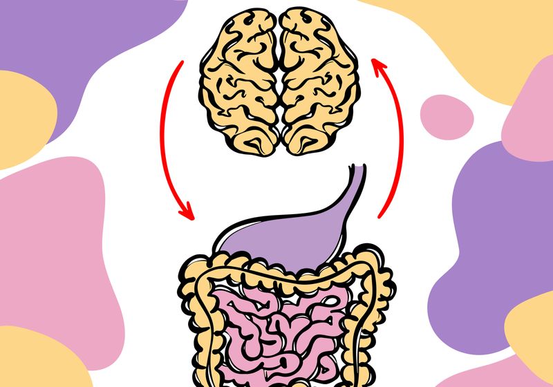
Immune cells can send signals between the gut-brain axis, but researchers found that these cells also migrate into the brain in a mouse model of Alzheimer’s disease.
The gut is home to a richly diverse community of microbes and nearly 80 percent of the body’s immune cells. This menagerie of gut-derived cells send signals along a bidirectional cellular highway, known as the vagus nerve, influencing not only the immune system but also brain function and behavior.
Due to this relationship, the gut-brain axis is emerging as a target in Alzheimer’s disease (AD). However, the immunological features of this axis in AD are not fully understood. This motivated researchers at the Buck Institute for Research on Aging to investigate and characterize the gut immune system in a mouse model of AD.
In a recent study, published in Cell Reports, the team observed that colonic immune cells migrated into the brain, likely mediated by a shared chemokine signal.1 In addition, a high-fiber dietary intervention could counteract these immune cell changes in the gut and alleviate AD-related pathology. Together, these findings provide further evidence for the importance of the gut-brain axis and the potential for dietary treatments to help reduce inflammation in AD.
To probe the intestinal immune landscape, the researchers examined colons from mice with and without AD. Using single-cell RNA sequencing, they found that mice with AD had upregulated neurodegenerative and immune inflammation pathways. Then, looking at the specific immune cell changes, they homed in on a B cell lineage cluster, which had a notable decrease in the amount of antibody-producing plasma cells.
To better understand the loss of B cells in the gut, the researchers examined the cells’ transcriptional features and saw signals linked to cell migration. The one that drew the team’s attention was the CXC chemokine receptor type 4 (CXCR4), which follows a chemical trail to subsequently bind to CXCL12. Indeed, the team found that the levels of CXCR4+ B cells decreased in the gut and increased in the brain, which correlated with higher expression of CXCL12 in the glial cells—inflammatory contributors in AD. Curious as to whether these B cells were linked to the gut, the researchers found that these were IgA-producing B cells. IgA contributes to intestinal homeostasis by neutralizing pathogens and promoting commensal bacterial colonization. These IgA+ cells accumulated in the brains of mice with AD.
Based on this observation, the researchers speculate that AD-related inflammation in the brain may recruit help from the gut immune system, which could weaken the colon’s cellular defenses and contribute to microbial changes associated with AD progression.
Given this migration of gut-specific B cells along the gut-brain axis, the team then hypothesized that blocking CXCR4 could prevent this movement. They tested this by injecting mice with a small molecule drug targeting CXCR4, which effectively inhibited the cells’ migration, leading to higher levels of CXCR4+ B cells and gut-specific IgA+ cells in the gut.
Finally, the researchers explored whether an anti-inflammatory fiber diet could provide benefits. Feeding AD mice inulin, a soluble fiber, for 13 months resulted in improved gut microbial balance, reduced brain neuroinflammation, and an increase in gut IgA+ cells. Notably, these mice also experienced a reduction in tremors.
“As far as we know, this is the deepest investigation of the gut immune system in a model of neurodegenerative disease,” said neuroscientist and coauthor Julie Andersen in a press release. “We look forward to studying its impact in other diseases including Parkinson’s and multiple sclerosis.”


